Proton Exchange Membrane Fuel Cells (PEMFCs) can become inefficient if the water cannot properly flow out of the cell and subsequently ‘floods’ the system. Until now, it has been very hard for engineers to understand the precise ways in which water drains, or pools, inside the fuel cells due to their very small size and very complex structures.
The solution created by the UNSW researchers allows for deep learning to create a detailed 3D model by utilizing a lower-resolution X-ray image of the cell, while extrapolating data from an accompanying high-res scan of a small sub-section of it.


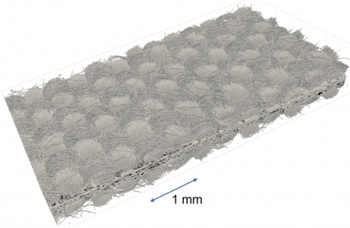
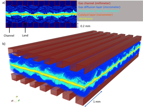
Top: 3D X-ray scan of a hydrogen fuel cell, showing carbon paper weaves, membrane and catalysts (in black). Scan provided by Dr Quentin Meyer. Bottom: 2D and 3D rendering of the segmented membrane electrode assembly with artificially overlayed flow channels. The gas channel and land contacting the GDL are labeled. Wang et al.
One of the reasons this research is so novel is that we are pushing the limit of what can be produced from imaging. It is very typical that when you use a piece of hardware, whether it’s a microscope or a CT scanner, the resolution of an image gets worse the more you zoom out.
Our machine learning technique resolves that problem, and the methodology is broadly applicable where any imaging is taking place, such as medical applications, or the oil and gas industry, or chemical engineering.
We have done preliminary super-resolution work with radiologists previously and we could surmise that by obtaining a higher resolution image from a larger field of view that it may be possible to diagnose diseases, such as tumour cells, earlier, when they are smaller.
—Professor Ryan Armstrong, co-corresponding author
The super-resolution algorithm, known as DualEDSR, improves the field of view by around 100 times compared to the high-res image.
During training and testing of DualEDSR, the algorithm achieved 97.3% accuracy when producing high-res modeling from low-res imagery. It also produced a high-resolution model in just 1 hour, compared to the 1188 hours (the equivalent of 50 days non-stop) it would have taken to obtain high-res images of the whole section of the fuel cell using a micro-CT scanner.
One limitation to the modeling process as detailed in the study is the fact the larger-scale low-res image and the smaller-scale higher-res image need to be taken at the same location, by the same machine.
These are known as Region of Interest scanners and are specialized pieces of equipment that may not currently be available at many facilities. However, the team hopes that further research will allow deep learning techniques to produce similar results in future when presented with images that were not taken at the same location and potentially not even using exactly the same instrument or material.